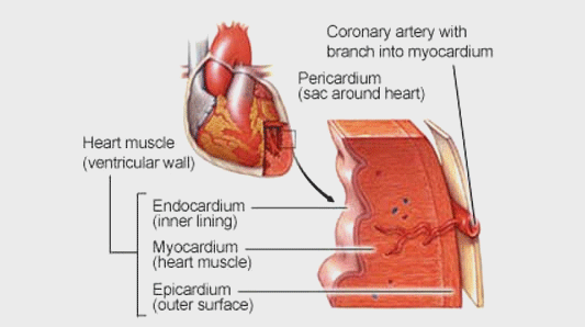
The wall of the heart has three layers: the outer epicardium
( epi = on, upon; cardia = heart), the middle myocardium ( myo = muscle), and the inner endocardium ( endo = within, inward).
The epicardium is the visceral layer of the pericardium. The majority of the heart is myocardium or cardiac muscle tissue.
The endocardium is a thin layer of endothelium that lines the chambers of the heart and the valves.
The pericardium is a triple-layered fluid-filled sac that surrounds the heart. The outer layer of this sac is the fibrous pericardium .
It is a strong layer of dense connective tissue. It adheres to the diaphragm inferiorly, and superiorly it is fused to the roots of the
great vessels that leave and enter the heart. The fibrous pericardium acts as a tough outer coat that holds the heart in place and keeps
it from overfilling with blood. Deep to the fibrous pericardium is the double layered serous pericardium. The serous pericardium is a
closed sac sandwiched between the fibrous pericardium and the heart. The outer layer is the parietal layer of the serous pericardium and
adheres to the inner surface of the fibrous pericardium. The parietal layer is continuous with the visceral layer of the serous pericardium
or epicardium, which lies on the heart and is considered a part of the heart wall.
Myocardium
The myocardium is the muscular wall of the heart and consists mainly of cardiac muscle. The myocardium's elongated, circular, and spiral
arranged networks of cardiac muscle cells, called bundles,
squeeze blood through the heart in the proper directions: inferiorly through the atria and superiorly through the ventricles.
Endocardium
The endocardium is an innermost, thin, smooth layer of epithelial tissue that lines the inner surface of the heart chambers and valves.
Cardiac Cycle
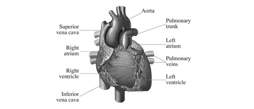
Blood flows through the heart in one direction only. It is prevented from backing up by a series of valves at various openings:
the tricuspid valve between the right atrium and right ventricle; the bicuspid, or mitral valve between the left atrium and left ventricle;
and the semilunar valves in the aorta and the pulmonary artery.
Each heartbeat, or cardiac cycle, is divided into two phases. In the first phase, a short period of ventricular contraction known as the systole,
the tricuspid and mitral valves snap shut, producing the familiar "lub"; sound heard in the physician's stethoscope. In the second phase, a slightly
longer period of ventricular relaxation known as the diastole, the pulmonary and aortic valves close up, producing the characteristic "dub"; sound.
Both sides of the heart contract, empty, relax, and fill simultaneously; therefore, only one systole and one diastole are felt.
The normal heart has a rate of 72 beats per minute, but in infants the rate may be as high as 120 beats, and in children about 90 beats per minute.
Each heartbeat is stimulated by an electrical impulse that originates in a small strip of heart tissue known as the sinoatrial (S-A) node, or pacemaker.
Blood
The fluid pumped by the heart that circulates throughout the body via the arteries,
veins, and capillaries. An adult male of average size normally has about 6 quarts (5.6 liters) of blood. The blood carries oxygen and nutrients to
the body tissues and removes carbon dioxide and other wastes. The colorless fluid of the blood, or plasma,
carries the red and white blood cells, platelets, waste products, and various other cells and substances.
Blood Vessels
The blood vessels are elastic tubular canals through which blood circulates in the body.
Different types of blood vessels include Arteries, veins and Capillaries. The blood vessels are part of the circulatory system and function to transport
blood throughout the body. The most important types, arteries and veins, are so termed because they carry blood away from or towards the heart,
respectively. The arteries carry blood away from the heart; the main arterial vessel, the aorta, branches into smaller arteries, which in turn branch
repeatedly into still smaller vessels and reach all parts of the body. Within the body tissues, the vessels are microscopic capillaries through which
gas and nutrient exchange occurs. Blood leaving the tissue capillaries enters converging vessels, the veins, to return to the heart and lungs.
Blood Pressure
The pressure exerted by the blood against the walls of the blood vessels,
especially the arteries. It varies with the strength of the heartbeat, the elasticity of the arterial walls, the volume and viscosity of the blood,
and a person's health, age, and physical condition. The pressure waves (pulse) can be felt at the wrist and at other points where arteries lie near the
surface of the body. Since the heart can pump blood into the large arteries more quickly than it can be absorbed and released by the tiny arterioles and
capillaries, considerable inner pressure always exists in the arteries. The contraction of the heart (systole) causes the blood pressure to rise to its
highest point, and relaxation of the heart (diastole) brings the
pressure down to its lowest point. Normal ranges for blood pressure in adult humans are:
Systolic between 90 and 135 mmhg (12 to 18 kpa)
Diastolic between 50 and 90 mmhg (7 to 12 kpa)
Blood pressure is strongest in the aorta, where the blood leaves the heart.
It diminishes progressively in the smaller blood vessels and reaches its lowest point in the veins.